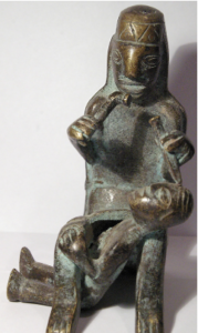
Bronze sculpture from Pisac, in the sacred valley of the Incas, Peru.
Inca’s were known to perform trepanations, however the exact purpose is unknown.
Trepanation, or craniotomy, is the act of making a hole in the skull to reach the brain. Skulls from many ancient cultures, including the Inca’s, show evidence that these civilizations successfully trephined themselves. Its apparent the patients survived these procedures, although we can only guess at the resultant cognitive state in primitive times, with primitive tools, and without much anesthesia.
Today, neurosurgeons use very sophisticated tools to perform craniotomies for many different conditions. The brain is nearly completely enclosed by the skull, but almost any area of the brain can be accessed. Using an endoscope, we at Rocky Mountain Brain & Spine Institute can remove a colloid cyst from the center of the brain, through a few centimeter incision and even smaller hole in the skull. Some patients can even leave the hospital the day following this type of procedure.
However, even using modern technology, there is some concern about the long-lasting cognitive effects of undergoing a craniotomy. There is a saying, “You ain’t never the same when the air hits your brain”. ~ Dr. Frank Vertosick Jr, MD
The brain is a unique bodily organ because it is not normally exposed to atmospheric pressure… the intact skull prevents atmospheric pressure from pressing in on all sides. The pressure around our brains (intracranial pressure- ICP) is normally less than 10cm of water relative to atmospheric pressure. When the skull is opened, atmospheric pressure equalizes with the intracranial pressure, and the full effect of this is not known.
We once planned brain surgery just to steer clear of “major” control centers. There are primary motor areas, sensory areas, vision areas, speech areas, etc… which surgeons avoid to prevent major and immediately noticeable postoperative deficits. However we are now much more aware of the “subtle” nerve pathways weaving throughout all areas of the brain, which when damaged by surgery, potentially could cause subtle and less apparent cognitive changes.
Joe Biden, for example, underwent two craniotomies for two aneurysms in 1988. Mr. Biden has given interviews as to his concern before pursuing these procedures. He remembered asking the neurosurgeon, “What are my chances of getting off this table completely normal?”. The surgeon reportedly replied, “35-50%… Well, your chances of living are a lot better”.
Newer studies have objectively looked at the potential cognitive impairments from craniotomies. Regarding the repair of aneurysms, postsurgical patients appear to perform below the norms on many neuropsychologist tests, although significant functional impairments are only seen in a minority of patients. And some of the changes may be more attributable to the aneurysm and/or rupture itself, as opposed to the surgical treatment. In studies assessing cognitive changes following craniotomy for tumors, again it appears that the tumor itself may have a greater detriment than the surgical procedure on postoperative cognitive changes.
In summary, advanced brain mapping and surgical techniques of the 21st century allow surgeons to tailor the craniotomy to limit the side effects of the surgery itself. However, neurocognitive changes do occur. These changes may be attributable to having a brain condition, and not necessarily from the surgery itself.


